


Acquired Grynfelt-Lesshaft hernia in a teenager patient
Universidad de Ciencias Médicas de Granma
Facultad de Ciencias Médicas Celia Sánchez Manduley
Manzanillo-Granma
VI Jornada Nacional de Clínica Virtual y 1ra Jornada Nacional
Científica Estudiantil de Presentación de Casos de Medicina Interna “Prof. Reinaldo Roca
Goderich in Memoriam”
Acquired Grynfelt-Lesshaft hernia in a teenager patient
Alejandro Macías Muñoz1
Jesús Daniel de la Rosa Santana2
Giselle Lucila Vázquez Gutiérrez3
Jimmy Javier Calás Torres4
1Especialista de I Grado en Pediatría. Asistente.
2Residente de Primer Año en Medicina General Integral
3Especialista de II Grado en Pediatría. Asistente. Investigadora Agregada
4Estudiante de 2do año de medicina. Alumno Ayudante de Inmunología
2021
“Año 63 de la Revolución”
ABSTRACT
Introduction: The lumbar hernia (LH) is a hernia rarely encountered in teenager patients. There is an increased incidence of traumatic aetiology of LH related to new diagnostic methods. LH have been frequently misdiagnosed as other surgical entities. It is presented a case of superior primary acquired LH in a teenager patient with no previous history of diseases.
Case description: a 14-year-old african-american male patient who complained of an occasionally painful swelling over the left side of the lumbar region, was clinically diagnosed with a Grynfelt-Lesshaft hernia and confirmed by CT scan. The patient was surgically intervened with a transverse lumpectomy incision over the tumour. The contents were reduced, and the 1cm x 1cm ring with no sac was closed and reinforced with polyester fibre prosthetic mesh. No immediate complications were observed and the patient was released of the institution five days after the procedure.
Conclusions: The Grynfelt-Lesshaft hernia is an uncommon surgical condition related to a congenital or acquired aetiology. The advances in diagnostic methods available allow to easily identify a Grynfelt-Lesshaft hernia in younger patients.
Keywords: hernia; surgery; pediatrics; adolescent; Surgical Procedures, Operative.
INTRODUCTION
The «lumbar hernia (LH) is a posterolateral body wall hernia where the bowel, omentum, or pre-peritoneal fat herniates through the lumbar triangles». (1)
It was initially described by P. Barbette in the year 1672 and later on R.J.C. Garangeot published the first case in 1731. (2) The incidence is most frequent between males of 50 and 70 years old. The LH is currently located over the left side on the lumbar region. (1) Most of the LH are primary acquired hernias, however the defect may be congenital or acquired. (3)
Moreno-Egea (4) et al. created a therapeutical classification system to identify the four types of LH. This classification has six criterion ordered in: size, location, contents, muscular atrophy, origin, and the existence of the previous recurrence.
LHs can occur through two well-defined areas in the lumbar region: the inferior lumbar triangle (Petit’s triangle), and the superior lumbar triangle (Grynfelt-Lesshaft’s triangle). (5) The «Grynfeltt-Lesshaft triangle is bound laterally by the inferior oblique muscle, the floor of the transversalis fascia, the medial border of the quadratus lumborum, and superiorly by the 12th rib». (1-2,5-6)
There is an increased incidence of traumatic aetiology. The advances in the diagnostics methods available are a cause of more encountered LHs in current medical practice. (2) However they are been misdiagnosed as lipomas, muscle strains, fibromas, abscesses, and kidney tumours. Each misdiagnosed is a cause of increased morbidity. (1)
The LH may present in patients as an asymptomatic lumbar mass. A lumbar mass with back pain and a lumbar mass with non-significant abdominal symptoms are others forms of presentation. (3)
It is presented a case of superior primary acquired LH in a teenager patient with no previous history of diseases, this is one of the first time that is reported this medical issue in a patient under 20 years old.
CASE REPORT
A 14-year-old african-american male patient, belonging to the urban area, presented to the paediatric hospital with no previous history of diseases. He noticed a swelling, occasionally painful, located in the left side of the lumbar region. This swelling increased on active movements and disappeared on prone position (Image 1). The patient’s mother refers to have noticed this swelling 7 years ago and two years before the consultation it was diagnosed as a lipoma by his family doctor.
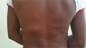
Figure 1
On physical examination, it was found a 7 cm x 7 cm mass over the lumbar area. The soft mass reduced when manually compressed and protruded painfully when the patient coughed. The mass was located horizontally between the 10th and 11th rib, and vertically between the paraspinal muscles and the external oblique muscle.
An X-ray was performed in search of fractures or abnormality at this level which was not found. The CT scan revealed the existence of a superior LH over the left side (Figure 2). The laboratory analysis performed did not show any relevant results.
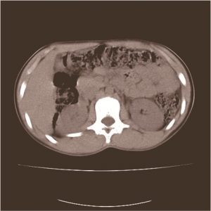
Figure 2
The patient was surgically intervened two months after the initial evaluation at consultation. The surgical approach was made with a transverse lumpectomy incision over the tumour, where a small ring of 1cm x 1cm containing the herniated tumour was found, composed of preperitoneal fat, no sac was described (Figure 3).
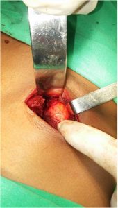
Figure 3
The contents were reduced, and the ring was closed by approximating its edges with independent points of assufil 2.0 prolonged absorption reinforced with polyester fibre prosthetic mesh fixed with U points of Prolene 2.0 (Figure 4).
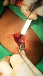
Figure 4
Penrose drainage was placed and independent stitches ensure the closure of the plans up to the skin. No immediate postoperative complications were observed. The patient was released of the institution five days after the procedure. A year after the surgery, the patient was consulted again denying any suggestive symptoms of LH and there were no tenderness or recurrence on physical examination.
DISCUSSION
Patients may refer low backache or a painful area over the herniated tumor. The palpatory finding and the swelling that dissapears in prone position is a current finding on Lhs (7) as in this case. The patient presented to the outpatient clinic didn’t have an emergency condition; this is due to most of the patients present to the health centers with non-emergency conditions and only 9% present as surgical emergencies. (2)
LHs are classified as congenital or acquired. An acquired LH is a primary or a secondary hernia according to the case. Secondary LH may be of traumatic or post-surgical aetiology, including flank incisions, renal surgery and other conditions. The estimation of secundary LH reaches the 25% of all acquired hernias. (7)
This diagnostic corresponds with a primary acquired LH that was initially described as a lipoma, this fact may be related with a misdiagnose of a LH as a lipoma or other medical conditions. (7) It is necessary to refer any cases of superior lumbar mass to a secondary level of attention and perform a deeper evaluation. As referred in the literature X-rays are not the imaging test of choice when diagnosing a LH. CT scan is the principal method for the diagnosis of a LH. (2)
Surgical repair of the hernia is the ideal choice. The classic technique is as good as the laparoscopic technique, although the second procedure will produce better outcomes. There is no real consensus on the best repair of a LH due to its low incidence (8) synthetic meshplasty is the most used of open repairs combined with muscle flaps, (7) this case only required the use of synthetic meshplasty for repairing the defect as there were no others issues such as a herniated sac with abdominal organs in its interior or a bigger ring that needed muscle flaps.
The open surgical technique proved to be effective a year after the surgery, younger patients have increased levels of tissue formation, (9) although younger patients are more active, this may affect the recurrence of LH or postoperative complications. The operated patient followed a strict schedule in the postoperative stages that did not involve the practice of intense exercises during the recovery, this fact helped to reduce the probability of recurrence.
CONCLUSIONS
The Grynfelt-Lesshaft hernia is a rare surgical condition produced by a congenital or acquired aetiology. A low backache or a located point of pain over the hernia in addition to a lumbar mass that reduces in prone position suggests a diagnosis of LH. The advances in diagnostic methods available allow to easily identify a Grynfelt-Lesshaft hernia in younger patients.
REFERENCES
- Suh Yiji, Gandhi Jason, Zaidi Saher, Smith Noel L., Tan Min-Yi, Khan-Ali Sardar. Lumbar hernia: A commonly misevaluated condition of the bilateral costoiliac spaces. Translational Research in Anatomy. 2017;8-9(September-November 2017):1-5. https://doi.org/10.1016/j.tria.2017.10.002
- Walgamage B. Thilan, Ramesh B.S., Alsawafi Yaqoob. Case report and review of lumbar hernia. International Journal of Surgery Case Reports. 2015;6(2015):230-232. https://doi.org/10.1016/j.ijscr.2014.07.022
- Zadeh R. Jonathan, Buicko L. Jessica, Patel Chetan, Kozol Robert, Lopez-Viego Miguel. Grynfeltt Hernia: A Deceptive Lumbar Mass with a Lipoma-Like Presentation. Case Reports in Surgery. 2015;2015:954804. http://dx.doi.org/10.1155/2015/954804
- Moreno-Egea A, Baena EG, Calle MC, Martinez JAT, Albasini JLA: Controversies in the current management of lumbar hernias. Arch Surg. 2007;142:82-88 . https://doi.org/10.1001/archsurg.142.1.82
- Ploneda-Valencia C.F., Cordero-Estrada E., Castañeda-González L.G., Sainz-Escarrega V.H., Varela-Muñoz O., de la Cerda-Trujillo L.F., et al. Grynfelt-Lesshaft hernia a case report and review of the literature. Anals of Medicine and Surgery. 2017; 2017(May2016):104-106. https://doi.org/10.1016/j.amsu.2016.04.002
- Matzke G, Espil G, Dos Ramos Alferes JP, Larrañaga N, Oyarzún A, Kozima S. Un recorrido por la pared abdominal: evaluación de las hernias por tomografía computada multidetector. Rev Argent Radiol. 2017;81(1):39-49. https://dx.doi.org/10.1016/j.rard.2016.04.009
- Sundaramurthy S, Suresh H.B., Anirudh A.V., Prakash Rozario A. Primary lumbar hernia: A rarely encountered hernia. International Journal of Surgery Case Reports. 2016; (20)53-56. https://dx.doi.org/10.1016/j.ijscr.2015.09.041
- Tchoungui Ritz FJ, Kouam V, Titcheu F. Primary Jean Louis Petit and Grynfeltt-Lesshaft concomitant hernias: A case report. International Journal of Surgery Case Reports. 2018;51(2018):1-4. https://doi.org/10.1016/j.ijscr.2018.07.040
- Guo S, DiPietro LA.Factors Affecting Woung Healing. J Dent Res. 2009;89(3):219-229. https://doi.org/10.1177/0022034509359125